Curcio, C. A., Allen, K. A., 1990. Topography of ganglion cells in
human retina. J Comp Neurol 300, 5-25. PMID 2229487.
Contact information, corresponding author: Christine A. Curcio:
e-mail
,
information
Click below for a copy of this article, selected figures, and associated
data.
Please be aware that the copyright is held by the publisher of Journal
of Comparative Neurology,
A.R. Liss
and that you can contact them for permission to reprint at
Wiley Online
-
Full text of the paper (searchable PDF)
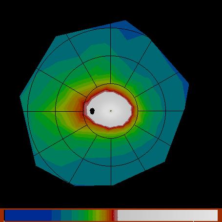
- Figure 5A:
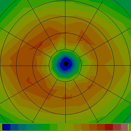
- Figure 5C:
- Text file
containing data representing ganglion cell density in a
composite eye, giving the co-latitude (in degrees), longitude (in
degrees), and density (in cells/mm^2). There are 171 points, followed by
330 triangles. The triangles are sorted by distance from the center of
the fovea. The Header says this is a Left eye, the radius of the globe
is 11.459 mm, the fovea is at 0,0 and the optic disc is at 20 degrees
co-latitude and 180 degrees longitude)
- Text file
containing data representing cone photoreceptor density in a composite
eye, giving the co-latitude (in degrees), longitude (in degrees), and
density (in cells/mm^2). There are 154 points, followed by 296
triangles. The triangles are sorted by distance from the center of the
fovea. The Header says this is a Left eye, the radius of the globe is
11.459 mm, the fovea is at 0,0 and the optic disc is at 20 degrees
co-latitude and 180 degrees longitude)
-
Fig. 6 Ganglion cell density as a function of eccentricity along
the horizontal and vertical meridians of the composite retina.
Last Modified: 8 January 2018
Kenneth Sloan